


Weight management
Research has shown that obesity quadruples the risk of CCL rupture. Weight loss decreases the wear
and tear on the abnormal joint and is an important aspect of non-surgical management. Weight loss alone may decrease the need for surgery in some obese patients. Being at a healthy weight can decrease systemic inflammation and the magnitude of force applied to joints. In dogs who have already torn one CCL, weight loss may delay rupture of the opposite CCL.
In one study, dogs with CCL disease that underwent a rigorous 12-week physical rehabilitation protocol with a rehabilitation specialist, followed by one year of home rehabilitation had an approximately 60% chance of ‘success’ with non-surgical management. Dogs who underwent the same rehabilitation protocol after surgery had a higher rate of success than those treated non-surgically, however if a dog is at a healthy weight at the time of diagnosis, medical management may have a much lower chance of success. +-
In one study, dogs with CCL disease that underwent a rigorous 12-week physical rehabilitation protocol with a rehabilitation specialist, followed by one year of home rehabilitation had an approximately 60% chance of ‘success’ with non-surgical management. Dogs who underwent the same rehabilitation protocol after surgery had a higher rate of success than those treated non-surgically, however if a dog is at a healthy weight at the time of diagnosis, medical management may have a much lower chance of success. +-
Physical Rehabilitation
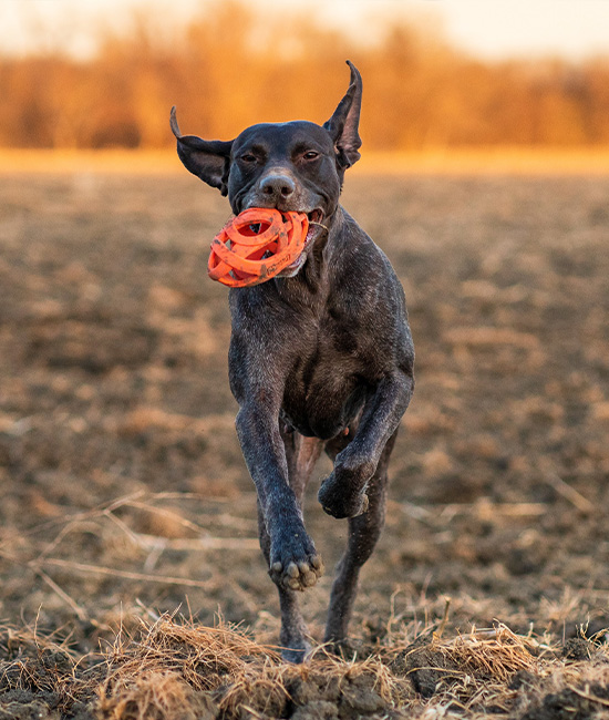
Regular, focused, controlled physical activity - particularly in a physical rehabilitation setting - can be
very beneficial to dogs with CCL rupture. Exercises such as stretching, range of motion exercises, controlled walking, underwater treadmill, and swimming, are supported in many scientific studies. Additional therapies such as Shockwave therapy and laser therapy can also be an important component of rehabilitation therapy for dogs with CCL rupture. At Midwest Veterinary Specialists, we have physical rehabilitation specialists and a state-of-the art rehabilitation center to maximize the outcome for your pet.
Physical rehabilitation offers many benefits, including:
+-
Physical rehabilitation offers many benefits, including:
- Improved limb use
- Weight loss
- Increased range of motion
- Increased muscle mass and strength
- Decreased pain
- Better mobility
- A happier, healthier pet!
Joint supplements and adjunct therapies
Unfortunately, there is little veterinary research to support or refute the broad use of joint supplements.
Joint supplements are unlikely to negatively affect your pet. If selected, it is important to choose high quality supplements that guarantee the amount of supplement in each pill and that have done studies to show that dogs can absorb them and that they affect the joints. There is evidence to support the use of diets supplemented with Omega-3 fatty acids and administration of polysulfated glycosaminoglycans.
+-
Omega-3 fatty acids

Fish-derived omega-3 fatty acids contain eicosapentaenoic acid (EPA) and docosahexaenoic acid
(DHA), and can benefit dogs. Several studies support the use of a diet rich in omega-3 fatty acids for dogs with osteoarthritis, demonstrating an improvement in weight-bearing, lameness, walking, and ability to rise from a resting position, and a decreased need for non-steroidal anti-inflammatory drugs (NSAIDs).
It is important to know that these products are not regulated by the FDA. It is therefore important to closely evaluate the content of the omega-3 supplement you purchase. Review the ingredients to ensure your dog will receive at least 20 mg/lb of EPA and DHA, and ensure you trust the brand you choose. +-
It is important to know that these products are not regulated by the FDA. It is therefore important to closely evaluate the content of the omega-3 supplement you purchase. Review the ingredients to ensure your dog will receive at least 20 mg/lb of EPA and DHA, and ensure you trust the brand you choose. +-
Polysulfated glycosaminoglycans
Polysulfated glycosaminoglycans (PSGAGs), inhibit enzymes that break down joint cartilage, and
inhibit production of chemical factors that lead to inflammation and pain. Adequan is one type of medication that contains PSGAGs and is administered as a series of injections over four weeks, and then on an as-needed basis.
+-
Platelet rich plasma (PRP) & Stem Cells
Other treatment options include injecting stem cells or platelet rich plasma (PRP) into stifle joints
affected by CCL disease.. A recent study evaluated the use of stem cells and PRP in patients with early partial CCL tears, finding significant improvement in some patients. Dr. Bergh has also had good success in treating canine athletes with early partial stable CCL tears with intra-articular PRP therapy and she is collecting data for a clinical study on this therapy.
+-
Pain management medications
CCL disease is painful and managing the pain often requires oral pain medications that may need to
be administered life-long.. Multiple medications may be combined for a synergistic effect to help keep your dog comfortable.
Nonsteroidal anti-inflammatory drugs (NSAIDs)
NSAIDs are one of the most commonly prescribed medications to treat pain in dogs. They are effective in blocking inflammatory and pain pathways, no matter where the pain or inflammation arises from. It is very important to note that while they are effective, they can have side effects that can potentially be serious.
These side effects can include:, vomiting, diarrhea, bleeding from the GI tract, inappetence, lethargy, and nausea. NSAIDs should not be used if certain concurrent disease processes are present, so please let our nurses know if your pet has any history of liver, kidney, or gastrointestinal problems or is on any other medications. If you observe any side effects after giving an NSAID, discontinue its use and contact us.
Gabapentin
Gabapentin is often used to treat neurologic conditions in people, and is also frequently used for pain control in dogs and cats. Gabapentin is especially good for controlling chronic pain and when used synergistically in a multimodal approach with other pain-relieving drugs. Gabapentin can cause sedation in some dogs.
Amantadine
Amantadine controls pain by blocking specific (i.e., N-methyl-d-aspartate [NMDA]) pain receptors. It has shown to successfully control pain when used in conjunction with NSAIDs. Side effects may include agitation and diarrhea. +-
Medical vs. Surgical Management
Medical management strategies should be comprehensive and multimodal for the best success. If there is significant instability present and/or tearing of the meniscus, it is much less likely to be successful. Surgical management consistently outperforms medical management and offers the most predictable long-term outcome.


Most commonly performed stabilization techniques
Extra-capsular techniques

Extracapsular techniques involve placing a synthetic material (e.g., suture or nylon line) around the
outside of the joint capsule in an effort to physically restrict and stabilize the joint , to allow scar tissue formation.
+-
- Lateral extracapsular technique — This technique uses a nylon suture line to provide stability around the outside of the joint (under the skin). By comparison to other techniques, this surgery is relatively inexpensive. Unfortunately, the nylon will stretch and loosen over time and the long-term stability is provided by scar tissue that forms along the path of the suture. As such, failure rates are higher in larger and more active dogs. If the repair fails and there is significant joint instability that causes pain and lameness, additional surgical or nonsurgical treatment is needed.
- Tightrope — The Tightrope technique uses a strong braided synthetic suture placed through bone tunnels in the femur and tibia. While the material resists stretching, it is more likely to cause infection and outcomes are not as consistent or favorable as with the classic lateral suture technique.
- Swivelock — The SwiveLock is similar to the Tightrope technique but relies on a bone anchor placed in the femur to secure the suture line.
Osteotomy techniques
Osteotomy techniques involve cutting the tibia bone to change the stifle anatomy. The goal of most of
the techniques is to reduce the tibial plateau slope/angle (TPA) from an average of 25 degrees to 5-6 degrees. This changes the joint biomechanics so that when a dog bears weight, the femur no longer slides down the tibial plateau. In essence, osteotomy procedures eliminate the need for the CCL without the need to physically restrain the joint.
Osteotomy techniques include:
+-
Osteotomy techniques include:
- Tibial plateau leveling osteotomy (TPLO)
- CORA-based leveling osteotomy (CBLO)
- Triple tibial osteotomy (TTO)
- Closing wedge osteotomy
- Tibial tuberosity advancement (TTA)

