TPLO Surgery
With a renowned board-certified surgeon.


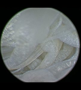
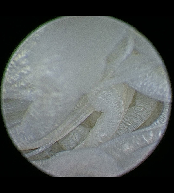
Anatomy of the canine stifle
Your dog’s stifles (i.e., knee joints) absorb a lot of impact as they run, jump, and play. To understand cranial cruciate ligament rupture, you must first understand the components of the stifle joint. This stifle is composed of the femur (i.e., thigh bone), tibia (i.e., shin bone), and patella (i.e., kneecap). Four ligaments—the cranial cruciate, caudal cruciate, medial collateral, and lateral collateral ligaments—are primarily responsible for stabilizing the stifle. The medial and lateral menisci act as cushions between the femur and tibia, and help provide congruency and further stabilize the joint.
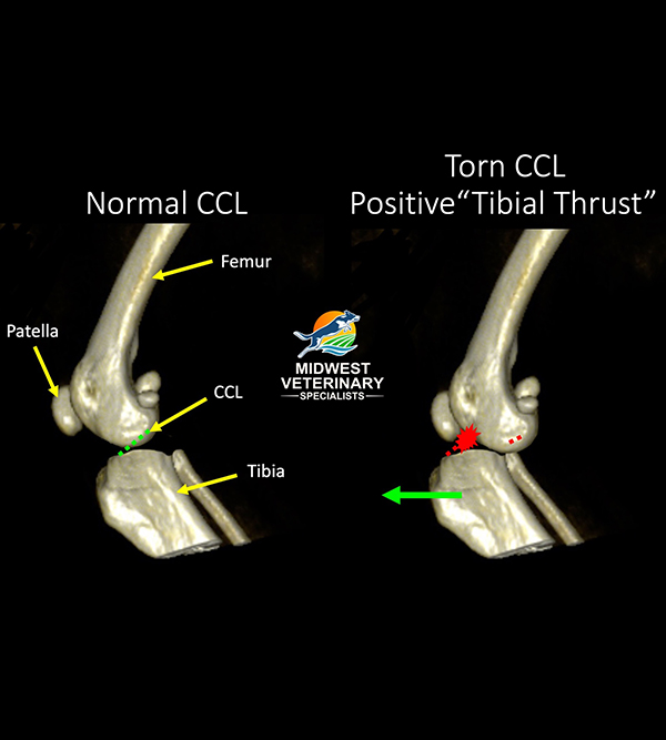
Canine cranial cruciate ligament
The canine cranial cruciate ligament (CCL), which is the anatomical equivalent to the human anterior cruciate ligament (ACL), has three functions:
- It limits hyperextension of the stifle
- It limits internal rotation of the tibia, relative to the femur
- It prevents front-to-back sliding motion (i.e., drawer motion) of the tibia relative to the femur.