A ruptured cranial cruciate ligament (CCL, sometimes called ACL) is the most common cause of hind limb lameness in dogs. If your dog ruptures their CCL, you want him or her back to normal activity level as soon as possible. Our team at Midwest Veterinary Specialists wants to help by offering information about CCL rupture and discussing what treatment options are available for this condition. We can also evaluate your pet and recommend the best treatment options for him or her!
Cranial cruciate ligament anatomy in dogs
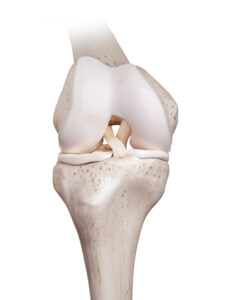
The CCL is one of two ligaments located deep inside the stifle (i.e., knee) joint that plays a very important role in stabilizing the joint. The knee is composed of three major bones: the femur (i.e., thigh bone), tibia (i.e., shin bone), and patella (i.e., kneecap). The joint is held together by numerous ligaments and the joint capsule. The medial and lateral menisci are thick cartilages that form a cushion and provide stability between the femur and tibia. The cranial and caudal cruciate ligaments cross over each other deep inside the joint (hence the name cruciate, or “crossing”), and prevent the tibia from slipping forward and backwards in relation to the femur.
Cranial cruciate ligament disease in dogs
CCL disease is a degenerative condition commonly caused by several contributing factors, including genetics, obesity, conformation, poor physical condition, and breed. Once the ligament starts to deteriorate, it is not able to heal and will continue to break down over time. Unfortunately, about 40% to 60% of dogs will ultimately develop CCL tears in both stifles over time. Two clinical pictures are seen in dogs affected by CCL disease. These include:
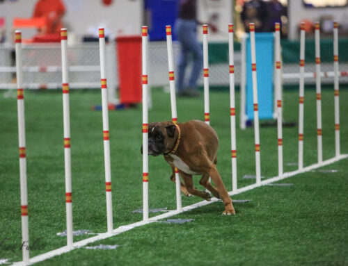
- Acute lameness that occurs during exercise — In most cases, the CCL has already been degenerating and a sudden large force causes the ligament to tear completely. In some cases the sudden and severe lameness may be due to tearing of the meniscus as well. Athletic large-breed dogs are at highest risk.
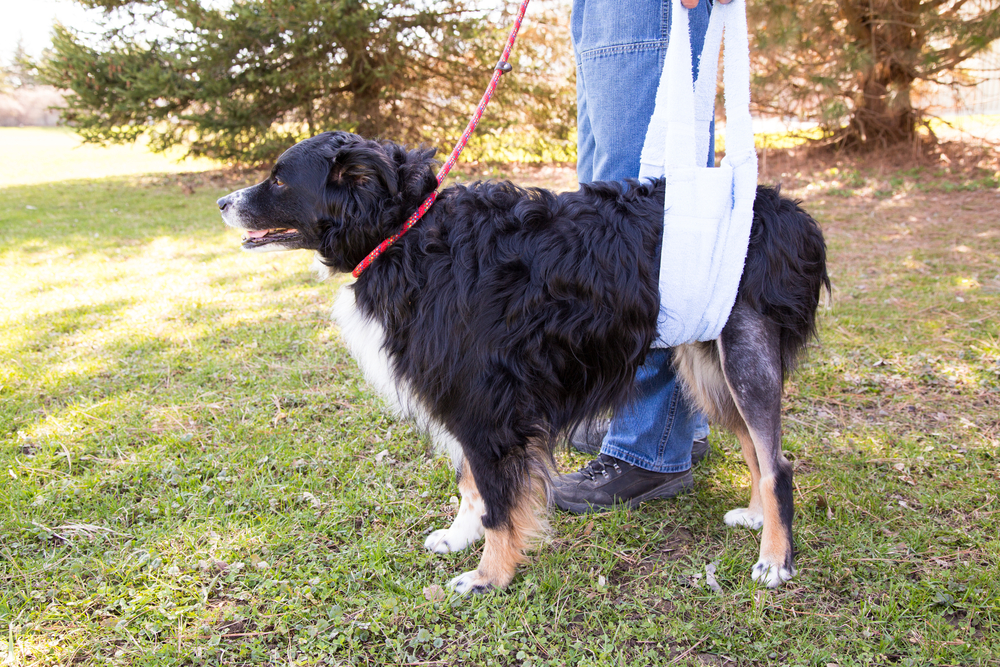
- Lameness that occurs gradually over time — In other cases, the CCL degenerates more slowly over time. These dogs may show lameness after heavy activity that improves with rest, difficulty rising after sitting, trouble jumping on surfaces, decreased overall activity, affected limb muscle loss, joint swelling, and overall stiffness.
While all dogs are susceptible to CCL disease, breeds at higher risk include rottweilers, Newfoundlands, Staffordshire terriers, mastiffs, Akitas, Saint Bernards, Chesapeake Bay retrievers, and Labrador retrievers. Small dogs frequently have CCL rupture in combination with medial patellar luxations.
Surgical treatment options for cranial cruciate ligament rupture
When the CCL is torn, the knee is unstable, causing sliding between the femur and tibia and subsequently potential damage to the menisci. Degenerative osteoarthritic changes occur, resulting in additional pain and loss of mobility. Surgery typically is recommended for dogs affected by CCL disease because this is the only way to effectively stabilize the joint to help prevent or minimize osteoarthritis. Surgery is also the only way to treat meniscal tears. All surgical procedures involve joint evaluation, which can be performed arthroscopically or through a larger arthrotomy. The techniques differ in how they eliminate the instability within the joint. Surgical outcomes are improved with experienced surgeons. The three most common surgical techniques used to treat CCL rupture are:
- Tibial plateau leveling osteotomy (TPLO) — TPLO is the best surgical option for stabilizing the joint after a dog ruptures their CCL. In fact, it is the only surgical procedure that has been shown to consistently allow dogs to return to normal function regardless of how big or small they are! The TPLO involves changing the biomechanics within the stifle joint so that the dog does not need the CCL any more. This is achieved by cutting the tibia, repositioning it, and holding it in place with a bone plate and screws. This procedure is complex and requires special training, but offers the best outcomes when performed by an experienced board-certified surgeon.
- Extracapsular repair (i.e., lateral suture)— This procedure involves using non-absorbable suture material to stabilize the knee joint. The suture is passed through a hole drilled in the front of the tibia and around a bone called the fabella. It runs roughly in the same direction as the CCL and acts as a physical restraint to stabilize the joint. Unfortunately, the suture will stretch or break post-operatively, and long-term stability relies on scar tissue that has formed along the suture path. If the suture breaks before enough scar tissue has formed, instability, pain and lameness returns.
- Tibial tuberosity advancement (TTA) — The TTA alters the biomechanics within the stifle joint by cutting the front portion of the tibia bone and advancing it forward. When the thigh muscles contract, instability is decreased within the joint. Good muscle strength and conditioning are needed for the best outcomes with this procedure.
After surgery, a dog’s activity levels typically are restricted for eight to 12 weeks to allow the bone and soft tissues to heal, and also to allow for re-conditioning. If post-surgery recommendations aren’t followed precisely, complications can develop. Rehabilitation therapy can help hasten and improve surgical recovery from surgery. At Midwest Veterinary Specialists, we have a dedicated rehabilitation service to help with rehabilitation following CCL surgery.
Non-surgical treatment options for cranial cruciate ligament rupture
Non-surgical therapies include pain management and physical rehabilitation therapy to build or maintain muscle and improve joint range of motion. Over time, scar tissue will form around the stifle joint and some instability will resolve, however the pain associated with meniscal tears will not subside with non-surgical management alone. Unfortunately recovery may take years and lameness often persists. At Midwest Veterinary Specialists, we have a team of veterinarians and nurses to help guide non-surgical management of CCL tears. Non-surgical treatments can include:
- Weight management — Lean and fit is the goal! Even a few pounds of weight loss can greatly decrease the wear and tear on your pet’s joints. If a pet is overweight, putting them on a safe weight loss program can greatly benefit by decreasing joint loads, increasing comfort, and improving mobility in dogs with CCL rupture.
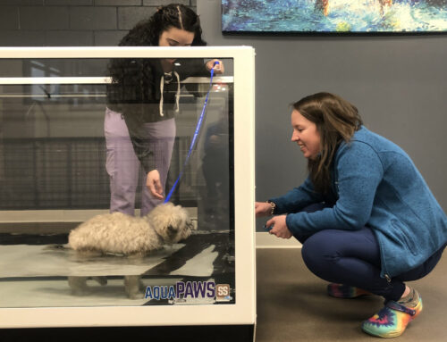
- Exercise moderation — Low impact activities such as leash walking and swimming are ideal. High impact activities such as running, jumping, or vigorous play should be avoided as they can increase pain, inflammation, and instability within the joint.
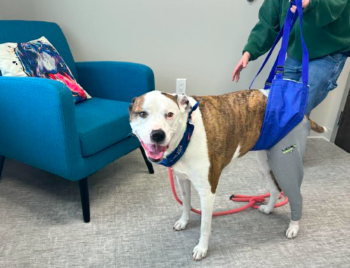
- Physical rehabilitation therapy — Formal physical rehabilitation therapy can help build muscle, improve range of motion, and aid in weight loss in dogs with CCL tears.
- Pharmaceuticals — Medications, such as non-steroidal anti-inflammatories, can help reduce pain and inflammation.
- Joint supplements — High quality joint supplements, which include glucosamine, chondroitin sulfate, ASUs, and fish oils can help maintain joint health and manage the osteoarthritis that goes hand-in-hand with CCL rupture.
- Joint injections — Dogs with partial CCL tears may benefit from joint injections with substances such as platelet rich plasma (PRP) or Hyaluronic acid (HA).
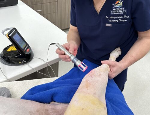
- Shockwave therapy — Sound waves can be used to stimulate tissue healing and decrease pain in dogs with CCL tears.
- Knee bracing — Custom knee braces are available, but unfortunately, they are not effective at stabilizing the instability that is present in dogs with CCL rupture and they can often cause sores or rubbing.
CCL disease can cause significant pain and mobility loss for your dog. If you notice your pet limping on a back leg, bring them in for evaluation as soon as possible. The earlier a problem is detected, the earlier treatment can be started, and the better the prognosis. Our team at Midwest Veterinary Specialists can determine what is causing the lameness, formulate a customized treatment plan, and get your pet back on all four feet as soon as possible.